Tuesday, January 31, 2017

Unit 6 Reflection

This unit was about biotechnology, the study and manipulation of living things to benefit mankind. Specifically, we learned about the domains of biotech, which are industrial and environmental biotech, agricultural biotech, medical or pharmaceutical biotech, and diagnostic research biotech, and examples of each domain. We also learned about some technologies in biotech, namely gel electrophoresis, polymerase chain reaction, and sequencing. Later, we learned about recombinant DNA technology, which creates transgenic organisms, and how to implement rDNA tech in the pGLO lab, an example of rDNA. Finally, we learned about morals, ethics, and values, and how these play a part in bioethics.


My strengths this unit were technologies of biotech and recombinant DNA. I found these topics pretty simple to grasp, as they were just processes that followed a logical sequence of steps in order to reach an end result. For me, that made them easy to understand. My weaknesses this unit were the domains of biotech, as I didn't really understand where the borders between domains lie, and bioethics, because ethics are not very logical and therefore harder to clearly understand. Also, I didn't have a lot of practice answering bioethical questions, so I'm also not great at those.

During this unit, we did two major labs: the Candy Electrophoresis lab, and the pGLO lab. From these labs, I learned more about electrophoresis, which is a technology involved with biotech, and how to transform bacteria with recombinant DNA, respectively. These labs helped me understand more concepts we learned during this unit.

Image result for recombinant dna

I would like to learn of more examples of each domain of biotech, so that I can not only learn about more practical applications of biotech, but I can also understand the distinctions between each domain. I would also like to learn about more technologies involved in biotech, as I found PCR, gel electrophoresis, and sequencing interesting and would like to learn about other similar processes. 

In my New Years Goals post, I said that my goals were to participate more in class and start studying earlier. Since we haven't had many class discussions, I haven't really participated in class discussions. However, we have had discussions in smaller groups, which I have done my best to participate in. Studying earlier hasn't been going as well. Instead of studying a week earlier for bigger tests, I've been studying the two days before.

Saturday, January 28, 2017

pGLO Lab Observations. Data Recording, and Analysis

1. Obtain your team plates. Observe your set of “+pGLO” plates under room light and with UV light. Record numbers of colonies and color of colonies. Fill in the table below.


2. What two new traits do your transformed bacteria have?


They are resistant to ampicillin and they glow in the presence of arabinose sugars.

3. Estimate how many bacteria were in the 100 uL of bacteria that you spread on each plate. Explain your logic.

I estimate that there were approximately 5,000 bacteria in the 100uL.

There were around 80 colonies in the +pGLO LB/amp plate. Since bacteria are very very small, I'd assume that there were a billion of them in one of each colony, making that plate have approximately 80 billion bacteria.

However, since there was LB on the plate, which bacteria feed off of, I think it is reasonable that there would be more bacteria at the end, as they would have been able to reproduce. However, since there was only 250 uL of LB in each plate, I think it is very possible that the bacteria ran out of food. For sake of simplicity, I assumed that the LB lasted for a day (24 hours) and the bacteria were able to reproduce once an hour.

Since bacteria reproduce exponentially, there were originally 80 billion divided by 2 to the 24th power, or approximately 5,000 bacteria.

4. What is the role of arabinose in the plates?

The arabinose triggers the green florescent protein, causing the bacteria to glow under UV light.

5. List and briefly explain three current uses for GFP (green fluorescent protein) in research or applied science.

One of the uses for GFP in research is bacterial transformation. The pGLO plasmid, which is transferred to bacteria, is a good example of bacterial transformation, as it is very easy to tell if the bacteria have received the pGLO plasmid. Using knowledge gained from this, scientists can do other experiments involving bacterial transformation.

Another use for GFP is that it could make specific cells glow. For example, GFP could be given to a certain cell, and then you would be able to see all the cells that had grown from that one, because they would also have inherited the GFP.

Finally, GFP can be used to genetically modify animals so they glow. As GFP has been shown to work on bacteria, it makes sense that GFP would also be able to work on other animals.

6. Give an example of another application of genetic engineering.

An example of genetic engineering is cloning. Cloning is a process that can create an organism that is identical to an already existing one through genetic engineering. Cloning could help save endangered species.

Tuesday, January 24, 2017

Bioethics Reading

Since I will be absent on Thursday (Jan 26), I have written a writeup on my bioethics reading instead of talking about it during class.

I read the article, UK approves three-person babies, by BBC News. As the article says, some people have genetic defects in their mitochondria. Since mitochondria are passed from mothers to their children, women with genetically defective mitochondria are unable to have healthy children.

The solution is a technique that combines a donor woman's healthy mitochondria with DNA from two parents. Either the parents' nuclei are removed from their embryo and placed into the donor embryo, or the mother's nucleus is removed from one of her eggs and placed into a donor egg. In both cases, the parents' embryo or egg is destroyed, as are the donors' nuclei. The resulting babies have 0.1% of the donor woman's DNA and can pass healthy mitochondria to their own children.

Some people believed that this procedure would bring hope to those with defective mitochondria, and would be safe and effective for all. Others questioned whether the babies were legal, voiced safety concerns, and suggested to wait for a safer procedure. It is estimated that 150 couples will be able to have babies using this technique each year.

Wednesday, January 18, 2017

Candy Electrophoresis Lab Focus Questions

When I analyzed the results of the gel, I noticed that the experimental samples had dye bands that were much larger than the dye bands of the known samples. This might signify that the experimental samples had many different dyes in them. The first experimental sample, which came from orange M&M's, turned into a long band that included both yellow 5 and yellow 6. The second experimental sample, which came from green Skittles, separated into a blue 1 band and a yellow 5 band, which makes sense, because blue and yellow make green. The last experimental sample, which came from red Mike and Ikes, was clearly red 40.


After looking at the structures of carminic acid, betanin, fast green FCF, and citrus red 2, and comparing them to blue 1, yellow 5, yellow 6, and red 40, I believe that fast green FCF would move similarly to blue 1, given their structural similarities. Also, I noticed that all the dyes in the example had negative charges, overall, and fast green FCF is the only one that has an overall negative charge.

Dog food manufacturers may put artificial food colors into dog food to make it look more appealing. Most people are drawn to brighter colors, so they might be more likely to buy brightly colored food.

Artificial food colors may be preferable to the natural food colors because brightly colored food is more attractive to the human eye.

The two factors that control the distance that the colored dye solutions migrate are the size of the dye molecules and the voltage of the gel electrophoresis system. Electrophoresis separates the molecules by size, so obviously, the size of the molecules of dye in each sample is important. However, the voltage is also important; the dyes would have moved faster if the voltage had been higher.

The force of the electricity would help move the dyes through the gel, as that is how electrophoresis works.

The fact that one side of the gel was charged positively while the other was charged negatively caused the molecules to separate by size. Since the dyes were negatively charged, they attempted to move towards the positively charged side. The speed at which they were able to do this was due to their size.

If I were to use gel electrophoresis on molecules with weights of 600, 1000, 2000, and 5000 daltons, I would expect the molecules with the higher molecular weights to move less far than the molecules with lower molecular weights. I would expect this because I think the molecules with higher weights would be larger, and therefore move slower.

Monday, January 9, 2017

New Year Goals

For this semester, I have two goals: to participate more in this class and to start studying earlier for tests.

In order to participate more, I will raise my hand to participate in biology class at least one time per class period when we are having a class discussion. If we are having discussions in our table groups, I will attempt to keep the conversation going by speaking multiple times.

In order to start studying earlier, I will begin studying for major tests approximately a week before the test date. I will begin studying for quizzes and similar minor tests half a week before. I will remind myself to study by setting reminders on my calendar and writing it in my planner.

Wednesday, December 14, 2016

Unit 5 Reflection

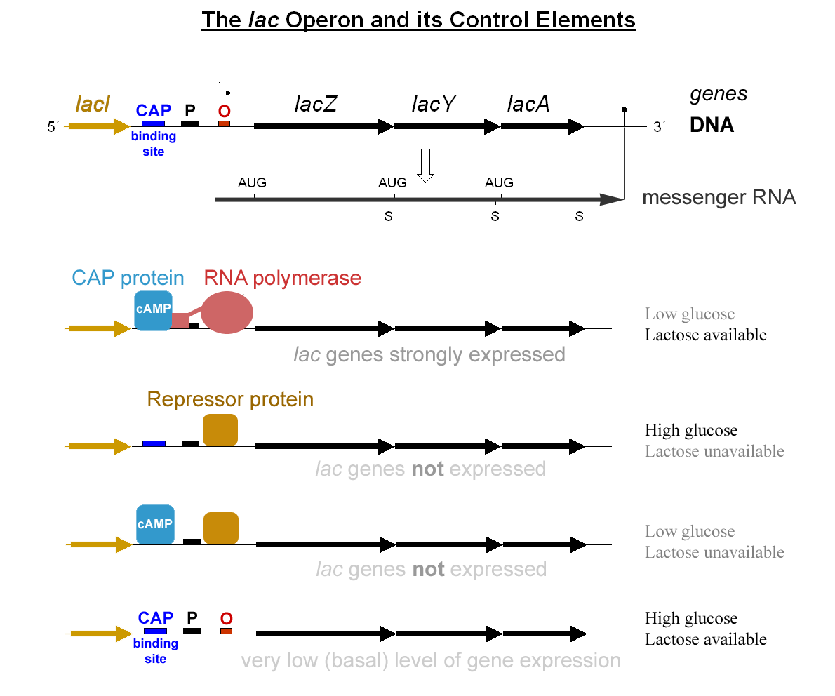
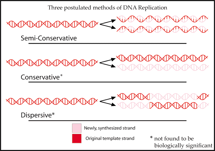
This unit was about the relation between DNA and physical traits. Some of the themes were the double helix, antiparallel shape of DNA; semi-conservative DNA replication, the process of creating two identical strands of DNA from a single one, each ending up with half of the original strand; transcription, how RNA is created in the nucleus; translation, how ribosomes read RNA to create proteins; the types of mutations: point mutations, frameshift mutations like insertion and deletion, inversion, and translocation; and how genes are regulated within an operon.


My strengths this unit were understanding semi-conservative DNA replication, transcription and translation, the different types of mutations, and the lac operon. I found these processes logical and easy to follow. My weaknesses this unit were understanding the antiparallel structure of DNA, doing transcription and translation by hand, and understanding DNA structure's influence of gene expression. I didn't understand antiparallel at first because it didn't make sense, but now I do. I struggled doing transcription and translation by hand because I am easily distracted. Originally, the topic of DNA structure and its influence on gene expression seemed very complicated, but now I realize it is pretty simple.

Image result for semiconservative dna replication

I would like to learn more about why DNA is shaped as it is, and why ribosomes can't just read DNA in the nucleus inside of the copied RNA. I am also curious about what would happen to the rest of the RNA if the ribosome was told to stop near the beginning of the RNA sequence. I would also like to learn about the probability for each mutation, and how efficient gene regulation truly is.

I am growing as a student by studying in a more effective way. I have begun creating flashcards and looking at more diagrams in order to study. This is an effective method to study for me because, according to the VARK Questionnaire, I have high visual, kinesthetic, and reading/writing scores. The flashcards are helpful because of my reading/writing score, while the diagrams are helpful due to my visual score. I have not yet found a way to incorporate my kinesthetic abilities into studying.

Monday, December 12, 2016

Protein Synthesis Lab Analysis

There are two main steps to make a protein: transcription and translation. In transcription, a section of DNA is copied, producing messenger RNA. In translation, the RNA reaches a ribosome. The ribosome reads bases 3 at a time. Each 3-base sequence corresponds to an amino acid, which the ribosome adds to the protein it is creating. After that, the amino acid chain folds and becomes a protein.

Image result for transcription and translation

There were three different types of mutations we tested: substitution, insertion, and deletion. Substitution either caused a great change or a very small change. Sometimes, it caused no change at all. Substitution was only an effective mutation when it was placed in a strategic position. Insertion and deletion both caused major changes when done at the beginning of a sequence. However, towards the end of a sequence, insertion and deletion caused much smaller changes.

Image result for substitution mutation

During this lab, I was allowed to choose a mutation of my own to test. I chose to test substitution, because after looking at the bases of the gene I was given, I realized I could easily substitute strategic nucleotides in order to get a drastically different result. I substituted two nucleotides in the beginning of the sequence, turning TAC into ATC. This changed the resulting RNA sequence from AUG to UAG. Basically, I changed the start codon to the stop codon. As you can imagine, the amino acids of the resulting protein were very much changed. Instead of the protein Met-Tyr-Lys-His-Val-Ile-Asn-Cys-Ile, there was no protein at all. This mutation changed more amino acids than any other tested. Obviously, placement did matter; if I just substituted bases at random, I would not have gotten the same result.


Mutations could affect our lives in many ways. An example of a mutation is Huntington disease. According to the U.S. National Library of Medicine's Genetic Home Reference, Huntington disease is caused by a mutation in the HTT gene, which provides instructions to make the huntingtin protein. Usually, in this gene, the sequence of cytosine, adenine, and guanine is repeated 10 to 35 times. However, within those with Huntington disease, this sequence is repeated much more, making the mutation an insertion. The length of the sequence of cytosine, adenine, and guanine creates very long huntingtin proteins, which are cut apart and bind together. This eventually causes neurons to die. Huntington disease is inherited with an autosomal dominant pattern.

Image result for autosomal dominant